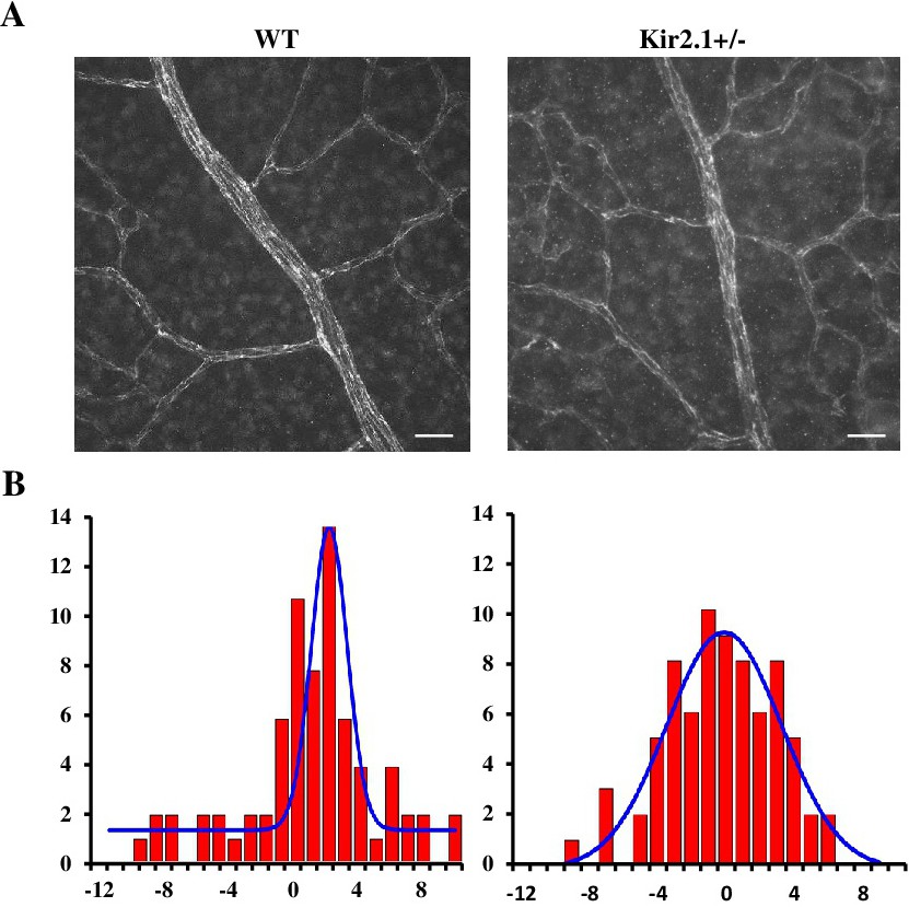
Fig. 5. Kir2.1+/- retinas show impaired endothelial cell alignment. Images of retina P6 stained for CD31 to visualize the contours endothelial cells. (B) Histograms of EC angles relative to the axis of the vessel determining the direction of the flow. Data are means ± SD of at least six mice per group. *P˂0.05. Scale bar panel A: 50 µm.